
Transforming Surgical Education with an Interactive Virtual Reality Resource Library
At the crossroads of medicine and cutting-edge technology, the Anatomy Laboratory Toward Visuospatial Surgical Innovation in Otolaryngology and Neurosurgery (ALT-VISION) at The Ohio State University is making transformative strides in surgical education. Leading the way with his 3D-models-turned-virtual-reality experience is Moataz Abouammo, MD, MSc, associate researcher and coordinator at ALT-VISION.
Abouammo collaborated with Je Beom Hong, MD, Ohio State Department of Neurosurgery research fellow, and Rebecca Leme Gallardo, MD, Ohio State Department of Otolaryngology research fellow.
The Exploration
Abouammo initially developed a comprehensive library containing more than 200 high-fidelity 3D models, simulating a wide range of endoscopic endonasal, transcranial and orbital surgical approaches. After rigorous surgical anatomical dissections, he used 3D photogrammetry — an advanced technique that compiles 3,000 to 4,000 photographs per specimen — to create precise, scalable reconstructions of surgical anatomy. These models were incorporated into the Atlas of Endoscopic Sinus and Ventral Skull Base Surgery.
Conceived as an interactive 3D dissection manual, the project aimed to provide detailed anatomical models for various endoscopic and skull base surgical approaches. But the vision extended beyond static models; the goal was to leverage technology to create a transformative educational tool that could enhance learning for trainees and practicing surgeons alike.
After Abouammo reached out to the ETI team, ETI Coordinator Mo Duncan and Learning and Development Specialist Thomas Ellsworth were able to troubleshoot and map out a path forward to take the 3D models even further.
The Solution
The collaborative effort resulted in scanning the previously developed 3D models and integrating them into a VR framework, crafting an immersive educational platform. Trainees and surgeons can now explore surgical approaches step by step, manipulate anatomical structures and visualize complex spatial relationships in a safe, interactive and three-dimensional space. This leap in educational technology bridged the gap between traditional dissection training and real-world surgical scenarios, enabling learners to rehearse procedures and build both confidence and precision without the risks associated with live surgery.
Looking forward, the team is poised to expand the project further. “Building on our success, we’re exploring ways to enhance the VR experience by layering the 3D models — allowing users to interactively peel back anatomical structures and visualize deeper surgical planes with precision,” said Abouammo. “We also plan to refine animations to better demonstrate critical steps in each approach, making the educational tool even more intuitive and dynamic. Furthermore, we are aiming to significantly expand the surgical scope to include maxillofacial, facial plastics, transoral and neck approaches.”
These advancements could revolutionize how surgeons train, offering a level of detail and interactivity that surpasses traditional methods. “With the ETI’s expertise in immersive technology and our shared commitment to innovation, I’m confident we can develop the next generation of surgical simulation tools, with potential applications in preoperative planning, interactive education for residents and medical students, patient education and beyond,” said Abouammo.

The Experience
Feedback from surgeons and trainees has been overwhelmingly positive. The immersive learning environment not only improved anatomical understanding but also increased trainees’ confidence and skill in complex procedures. Early results indicate that the tool is transforming surgical training by making learning more engaging, accessible, fun and effective. As long-term clinical impact is studied, the immediate educational benefits are clear — and the team looks forward to continuing their collaboration with the ETI to push the boundaries of what is possible in surgical education.
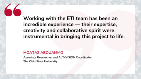
Abouammo shared the following: “Working with the ETI team has been an incredible experience — their expertise, creativity and collaborative spirit were instrumental in bringing this project to life. They were not only highly skilled in troubleshooting technical challenges but also deeply invested in the project’s success. They treated the project as if it were their own, constantly brainstorming ways to enhance its educational impact.
Thanks to their expertise, we were able to deliver a cutting-edge VR training tool that received outstanding feedback from surgeons and trainees alike. I’d jump at the opportunity to work with them again on future projects.”
Kyle K. VanKoevering, MD, FACS, associate professor, otolaryngology — head and neck surgery at Ohio State College of Medicine, praised Abouammo’s innovative thinking. "This novel, immersive technology allows learners to experience surgical skull base anatomy in a way that is unlike anything available,” said VanKoevering. “More portable and accessible than any cadaveric dissections, these 3D models have the potential to revolutionize anatomic education."

