Image
In an innovative effort to enhance the educational experience of her critical care nutrition course, Kristen Roberts, PhD, RD, LD, partnered with Laura Boucher, PhD, AT, ATC, to teach students on the path to becoming dietitians. The advanced knowledge of human anatomy that students glean from Boucher gives students a better understanding of muscle and fat variations that helps them master the physical assessment skills needed to obtain their credential and perform their job duties as clinical dietitians.
Initially, the collaboration between Roberts and Boucher, both clinical associate professors at The Ohio State University, involved a class of dietetics students watching Boucher teach with a prosected donor body. While both professors agree that nothing can replace the experience of seeing anatomical structures in a real body, there were drawbacks to the approach for this discipline. While some dietetics students were up front and asked a lot of questions during the dissection, many of them hadn’t had dissection experiences and weren’t comfortable engaging.
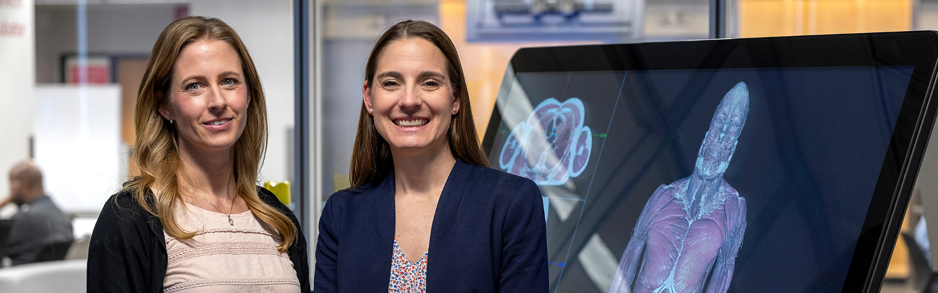
When the two reached out to ETI Coordinator Mo Duncan, Roberts and Boucher sought out the cutting-edge educational technology tools in the EdTech Incubator’s (ETI) Anatomy Visualization Zone. They were interested in replacing the donor prosection teaching with a 2D dissection on the Sectra table and 3D visualization of a full body in virtual reality (VR), with hopes that they could engage more students while decreasing costs compared to using a donor prosection.
The transition to the Anatomy Visualization Zone from the anatomy lab brought encouraging improvements to the learning experience. Technology used in the zone includes:
During the lab time, students were divided into three stations. One station included looking at the vascular system with Quest 3 headsets, and the second station was an interactive, virtual dissection led by Boucher. Boucher showed musculoskeletal anatomy, different cross-sections and thicknesses of fat, providing a deeper understanding of anatomical variations as students asked questions. She reported that more students felt comfortable being close to the virtual dissection and therefore more students were engaged in the learning experience.
“It worked really well,” said Boucher, “and we've had a lot of years of the donor body experience, but we want to come back to the [ETI] space. And I think that's a testament to the potential that's there and the feedback from the students.”
The third station involved students independently looking at the same body that Boucher showed on the Sectra table using the Varjo headset. Students could isolate structures and explore different parts of the body while others could watch the experience on a large TV screen.
Duncan provided guidance and training during two preparation sessions before students came in for their lab. There were a couple technical speedbumps to work through with the vascular system stations and Roberts and Boucher plan to cut out those stations the next time they use the space so students can get more time at Boucher’s virtual dissection station.
When asked about using the anatomy lab versus the Anatomy Visualization Zone, Roberts said, “I think there were trade-offs to both, but to me, the overarching positive with the [Anatomy Visualization] space was that I actually think their learning was enhanced because of how the time was spent.”
“I think this fall I'm going to try to go into the Anatomy Visualization area to take advantage of a similar set up with the MAT [Master of Athletic Training] students, because I think they might get more out of their general medical conditions course where we teach abdomen, thorax, pelvis and heart and lung anatomy,” says Boucher. “This experience has shown me what other opportunities there are and where I can apply the same type of learning in a class to have a better experience that might be more meaningful.”
![]()
EdTech Incubator
4th Floor, Prior Hall
376 W 10th Ave
Columbus, OH 43210
eti@osumc.edu
If you have a disability and experience difficulty accessing this content, please submit an email to ITDigitalAccessibility@osumc.edu for assistance.