Throughout 2024, a team of The Ohio State University College of Medicine faculty members and Health Sciences Library (HSL) staff worked together to launch the use of syGlass for the HSL’s EdTech Incubator (ETI), and we are excited to announce that syGlass has been approved for use!
syGlass is a sandbox virtual reality software with robust capabilities for immersive learning, streamlining research and improving patient care. SyGlass provides stereoscopic visualization that allows users to closely examine, manipulate and explore medical images in ways that they previously weren't able to. The software transforms the way physicians, students and medical professionals understand and interact with complex anatomical structures and diagnostic images, making syGlass a great example of the Wexner Medical Center’s “Reimagining Learning and Development” priority in its Impact 2035 Strategic Plan.
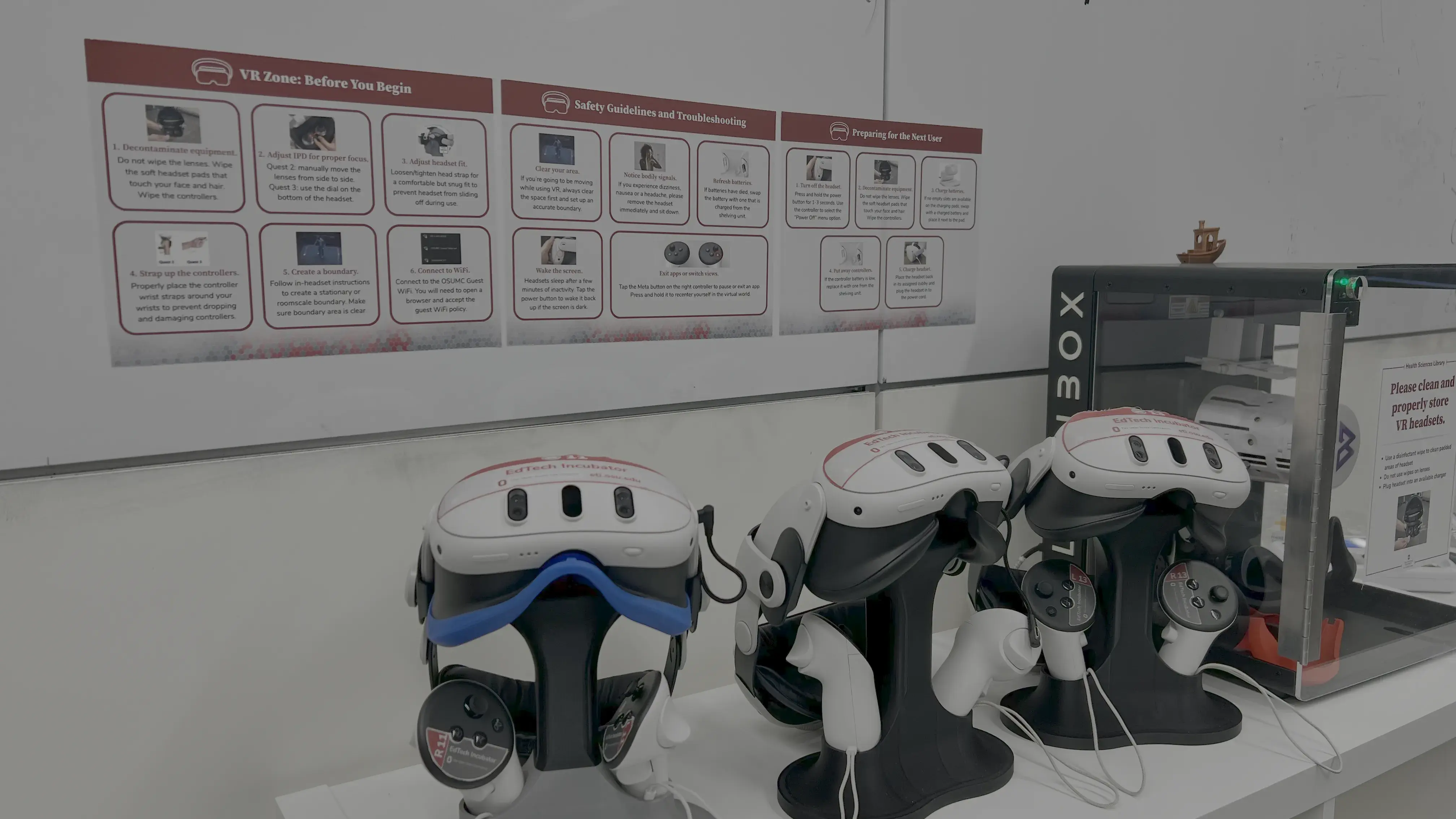
The ETI made a single-use purchase of the software, so individuals do not need their own subscriptions to come in and utilize the VR technology. Using syGlass is intuitive, with a low introductory period because the controls are made to feel natural to use. You don’t need to consider yourself a high-tech individual to use syGlass.
syGlass Applications
Clinical:
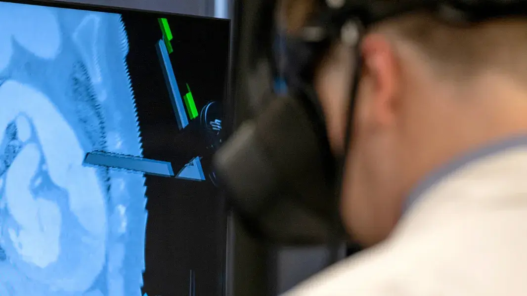
If physicians have consent from patients, they can transform a patient’s 2D scans into 3D images and examine them in virtual reality. This immersive exam of the scans reveals intricate patterns and relationships in patient scans that are difficult to recognize in traditional 2D imaging. Physicians can zoom into areas of interest, rotating and manipulating the images in 3D space to view them from different angles. This provides a much richer understanding of patient anatomy, which can be critical for diagnosis and treatment planning.
Educational:
Seeing patient scans in virtual reality provides students with better visualization and deeper understanding of what they can see and find on patient imaging. Faculty members can record videos of themselves manipulating the 3D images and splicing through layers of anatomy as they narrate what is shown on the scan. As students watch the recording with a VR headset, they can pause the video and take control of the 3D image to do their own examination, and when they press play it will go right back to the narrated video. A prime example of how syGlass is used for educational purposes comes from Dr. Blair Suter, a cardiologist at the Wexner Medical Center and College of Medicine faculty member. Suter is currently conducting a pilot study that uses a patient’s CT scan of their Watchman device, which is implanted in the heart to prevent strokes. Using syGlass, Suter recorded a video of himself manipulating the scan in 3D to show residents and fellows how the patient’s device was mal-positioned. Suter hopes to build a library of videos like this to give his fellows, residents and students the ability to explore many scenarios and complications – all in a risk-free environment that helps build their experience with cardiac imaging. Read more about Suter’s pilot study in this impact story: Cardiologist Blair Suter, MD Utilizes New VR Technology for EducationCardiologist Blair Suter, MD Utilizes New VR Technology for Education.
Research:
syGlass is a robust research tool for data visualization. Researchers can bring in multiple scans to track differences, with big data capabilities allowing for terabyte-size image files. When recording video notes or making dissertation presentations, users can pull up multiple scans at the same time to talk through the comparison. The software can also help with pattern recognition, cell counting, object tracing and volume segmentation.
Acquisition and Implementation Process
SyGlass use at the ETI resulted from Dr. Suter’s inquiry to use ETI technology for training residents and fellows in cardiac imaging. Initially, Suter looked into utilizing the 3D Printing Lab to translate 2D patient CT scans into 3D when Derek Harmon, PhD, Ohio State College of Anatomy associate professor – clinical, recommended using syGlass based on prototypes of the software he had seen.
The software was demoed by ETI staff and Ohio State physicians with Quest 3 and Varjo headsets. The ETI moved forward with the Varjo headsets for syGlass use because they could handle transforming bigger data sets into 3D with less visual delays. The headset itself also has fans to keep someone from overheating as they practice and record videos. In order to explore all uses of syGlass, including uploading patient scans, ETI staff worked closely with Ohio State and Wexner Medical Center IT teams to address processes for ensuring patient privacy and HIPAA compliance. syGlass even pushed updates to our demo software so the latest version could go through risk assessment with the IT team. syGlass is a breakthrough in clinical care and research, and transforms the future of medical education. It is preparing the next generation of healthcare professionals for the challenges of real-world medicine. This software is bridging the gap between theoretical knowledge and real-world application.
If you’re interested in using patient scans in syGlass, ETI Coordinator Mo Duncan can verify if your scans are compliant before using them in the VR software.
Ready to check out the software for yourself? Contact the ETI to explore syGlass.